Amazing Image Earns Entry Into International Contest
September 17, 2024 — Dr. Michel Becuwe has spent a good amount of his career examining life under a microscope.
An assistant professor of biology at Marist, Dr. Becuwe joined the College in 2022 after a stint as a course instructor in molecular and cellular biology at Harvard University. Born in France, he led his first summer attachment trip to Paris over the summer.
Dr. Michel Becuwe with Marist student Nicolas McGuire ’25 during an attachment course in Paris. Photo courtesy of Dr. Becuwe.
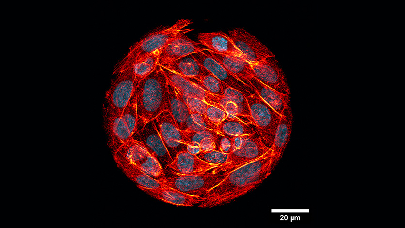
Dr. Becuwe is a finalist in the French Embassy’s contest, “An Artistic Odyssey Through Science,” for an image captured during a summer trip to Paris. The image shows Chinese hamster ovary cells in a circular pattern, illustrating how cells adapt their shape and organization to their environment.
It was taken using a confocal microscope, which uses lasers and pinhole that capture a high-resolution image at 200x magnification.
The imaging project was part of a collaboration between Marist students and their French counterparts at the Cergy Paris University.
“This course demonstrates how college connections can impact your life long-term,” said Dr. Becuwe. “We developed a collaborative workshop pairing Marist students with Cergy master's students to work on biology research projects using advanced microscopes. I'm thrilled that one of the images from this course was selected for a photo competition. The course will run every spring, and I’m excited for more stunning images and impactful discoveries in the future.”
Dr. Becuwe’s Marist students in Paris present the results of their research on the image. Photo courtesy of Dr. Becuwe.
The cells were stained using nuclear (blue) and cytoskeleton (orange) fluorescent markers, creating a stunning visual representation of cellular organization. The project was part of a two-week spring program, "Power of Microscopy in Biology Research," which allowed Marist students to gain hands-on experience in advanced biological imaging techniques.
How to Vote Online
Voting is open until October 15, and it’s quick and easy to cast your vote. Click HERE to vote and support Dr. Becuwe’s work!